Oil & Gas is a specific field of applied geology that combines economic and structural geology with methods of mineralogy, palaeontology and sedimentology. The mineralogical, structural and palaeontological capabilities of scanning electron microscopy provide a wide range of tools that are widely used in reservoir rock characterization.
The mineralogical aspects are based on Automated Mineralogy using the TIMA analyzer. TIMA provides information on the distribution of minerals and the pores in the reservoir rocks. This approach is most commonly applied to the “conventional” reservoir rocks formed by coarser-grained sediments. The important authigenic minerals lining the pores can be identified as well as the porosity itself. Image analysis tools are used to provide information on pore size and geometry and other features.
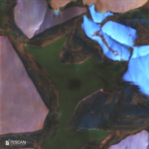
Quartz cement overgrowing detrital quartz grains imaged by cathodoluminescence
- Cathodoluminescence (CL) helps to identify authigenic overgrowths of minerals on the clastic grains e.g. quartz cement in quartz sandstones, which can block pores.
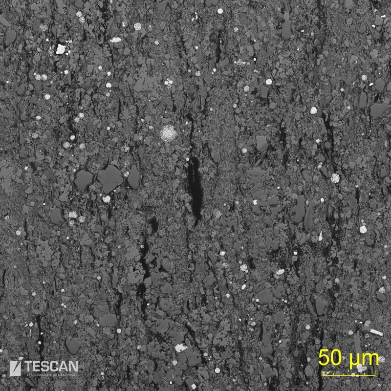
- The Automated Mineralogy method can also be used to characterize the “unconventional” reservoirs formed by fine-grained sediments such as shale.
- Modal mineralogy also helps in predicting the response to fracture stimulations.
- The textural analysis combined with the mineralogical composition is used to identify different sedimentary facies from both cores and from drill cuttings.
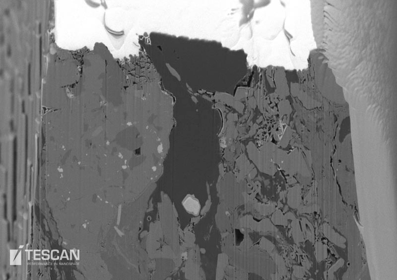
- The fine-grained un-conventional reservoirs are evaluated using advanced dual beam systems. In these systems, the focused ion beam (FIB) is first used to create a cross-section of the sample and the electron beam is then used for imaging the cut sample surface. Successive slices can then be used to build a three-dimensional image.
- The surface finish of each cross-section created by the FIB is very fine and can reveal nanometre-sized details. The FIB technique is thus the optimal tool for obtaining 3D reconstructions and permeability evaluation.
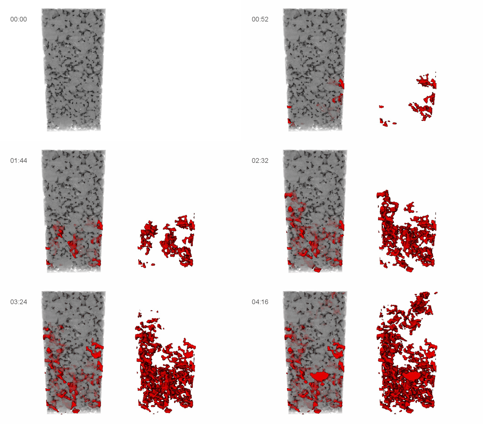
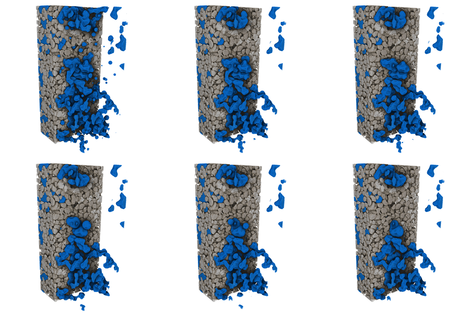
- The CoreTOM can be utilized to provide multi-scale micro-CT data on rock cores up to 1 meter in length and 300 mm diameter. Additionally, Dynamic micro-CT can be used to perform uninterrupted in situ experiments examining topics such as multi-phase flow and rock fracture.
- Section through a shale sample
- FIB cross-section of an oil shale sample ready for 3D data collection
- Visualization of oil displacing water
- Time lapse of CO2 dissolution


