The study of mineralogical and petrographic samples using scanning electron microscopy is now routine in geological research. Similar to optical microscopy but with a much higher resolution, electron microscopy reveals detailed textural relationships between the mineral grains. Its analytical capability has a much wider potential because the interaction between the electron beam and solid matter create several very useful output emissions. Among the most important emissions are backscattered electrons (BSE), secondary electrons (SE), characteristic X-rays and light photons.
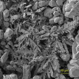
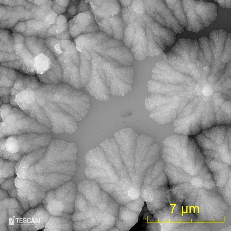
Dendritic crystals of native silver
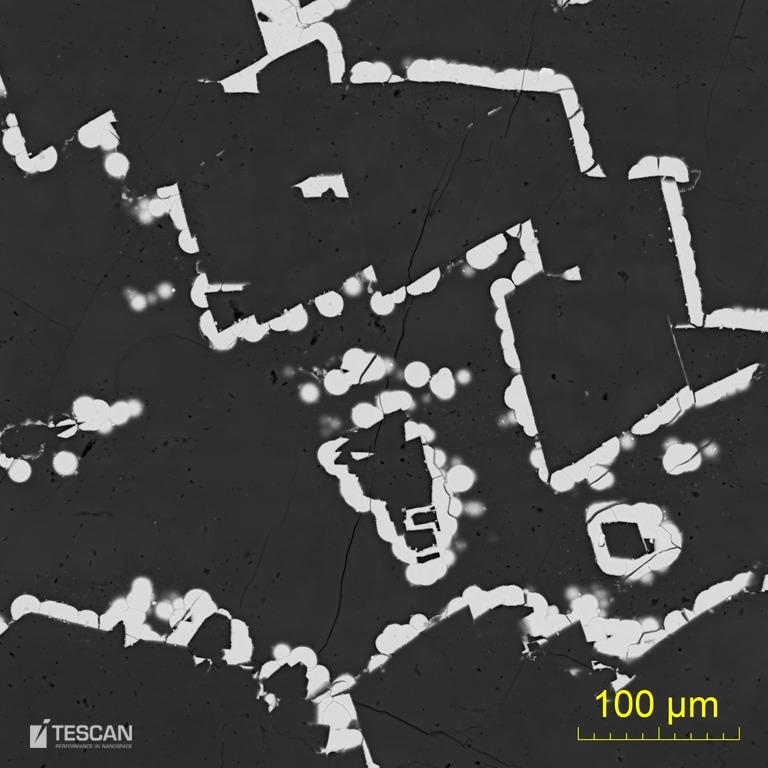
- The intensity of the Backscattered Electrons is directly proportional to the average atomic number of the observed phases and is used for imaging, distinguishing individual mineral grains and identifying zones of discrete phases. The contrast resolution can discriminate differences as low as about 0.1 atomic number. The backscattered electron signal enables the user to track zonation in mineral phases and to find optimal analysis points. Similarly, it can be used to visualize and locate specific phases containing heavy elements. This is especially important in searching for very rare but very valuable, phases that generally form small grains e.g. gold and platinum group minerals. Differences in the BSE intensity can also be used to identify the variable orientation of individual crystals of aggregates.
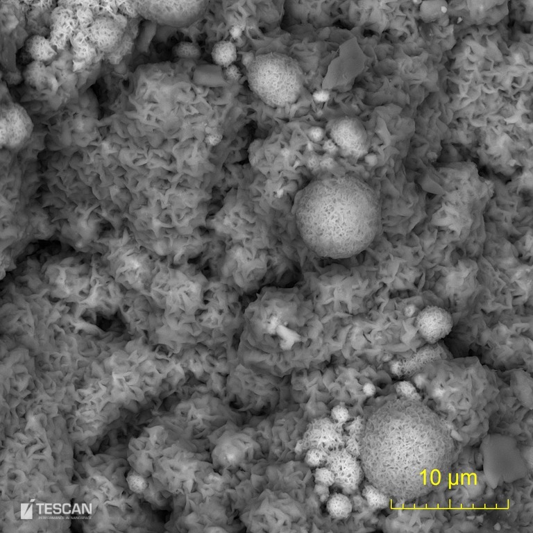
- Secondary Electrons are typically used to observe the morphology of three-dimensional samples. They are formed closer to the surface than BSE, have a high spatial resolution, a large depth of field and they are less sensitive to differences in the atomic number of the material.
- Characteristic X-rays are the most important output of the electron beam – solid matter interaction as they are used to identify the phases based on their chemical composition. This capability can be used for interactive qualitative assessment but most importantly for quantitative analysis. Special third party detectors compatible with the whole portfolio of TESCAN microscopes are used for this purpose.
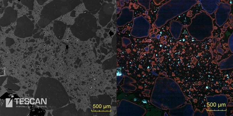
- Light Photons are also a useful product of the electron beam – sample interaction. This phenomenon is known as cathodoluminescence (CL) and produces many different luminescent phases among minerals. Many mineral species differ by the colour of the light emission. The colour CL can be used to distinguish different common rock-forming minerals e.g. feldspars or carbonates. CL is also very sensitive to differences in trace element composition and structural order of some minerals. These effects can be observed using panchromatic (black and white) or colour CL detectors. TESCAN manufactures panchromatic detectors and a four-channel combined panchromatic and colour detector – Rainbow CL. A combined solution for simultaneous acquisition of BSE and CL images is also available.
- Autunite crystal filling voids in goethite
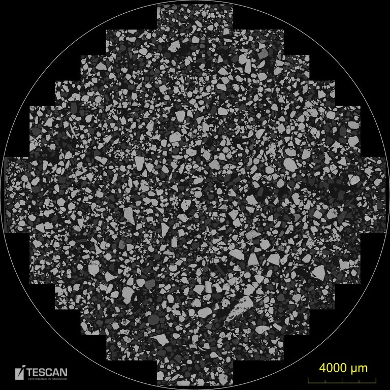
- Stitched image of heavy mineral sand (25 mm in diameter)
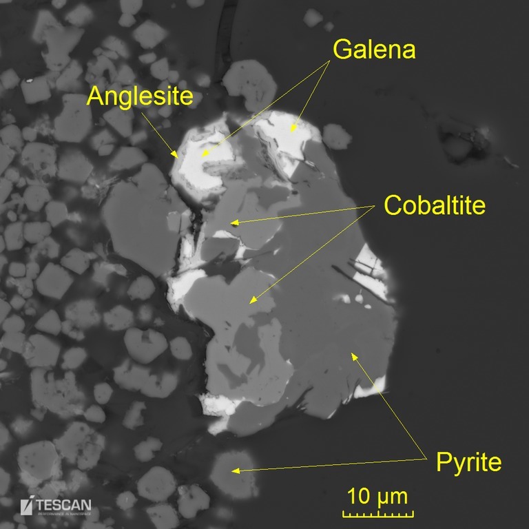
- Partially weathered sulphide grain accompanied by euhedral pyrite
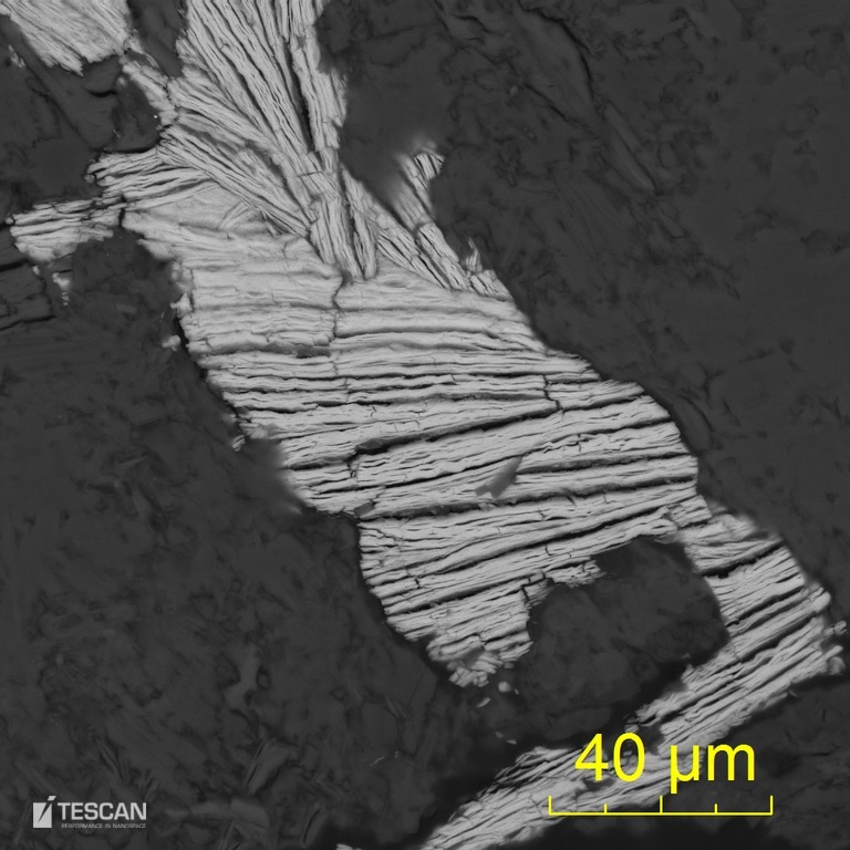
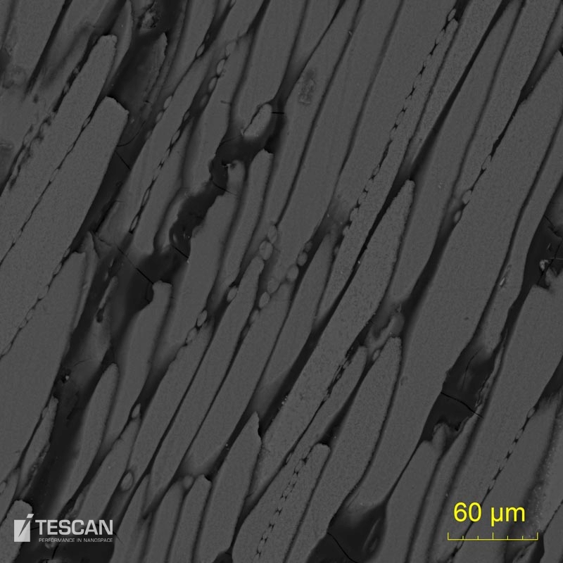
- Cookeite tabular crystals
- Iron oxide encrustation on a rock fracture
- Secondary copper phases formed by chalcopyrite weathering
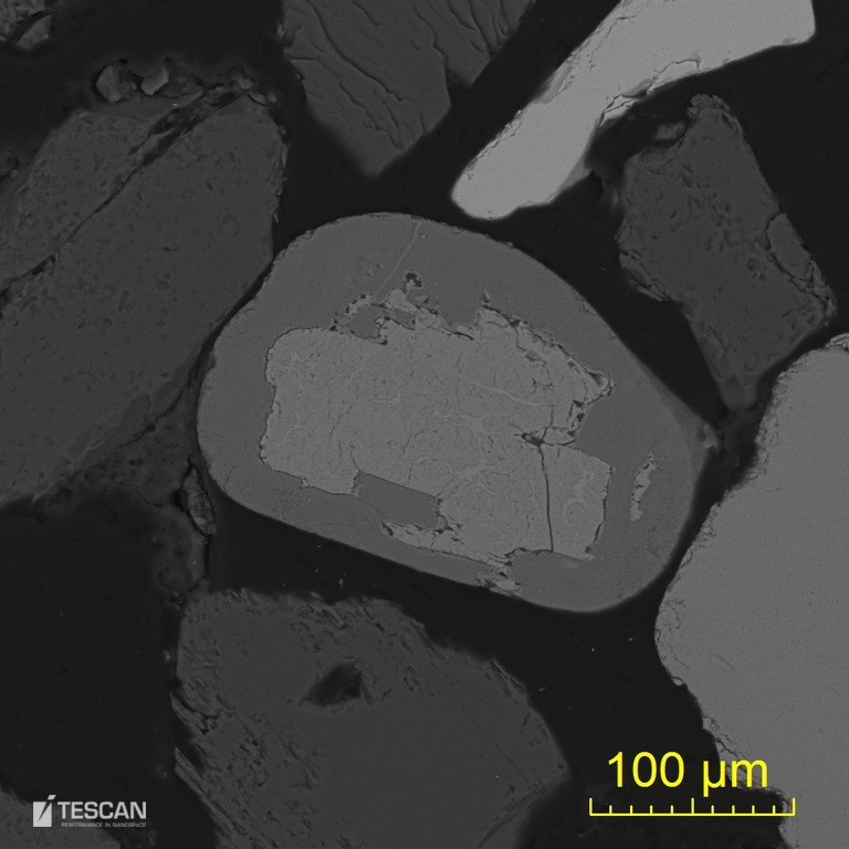
- Ilmenite grain which underwent partial leucoxenitization
- Uraninite crust (light) in calcite gangue (dark)
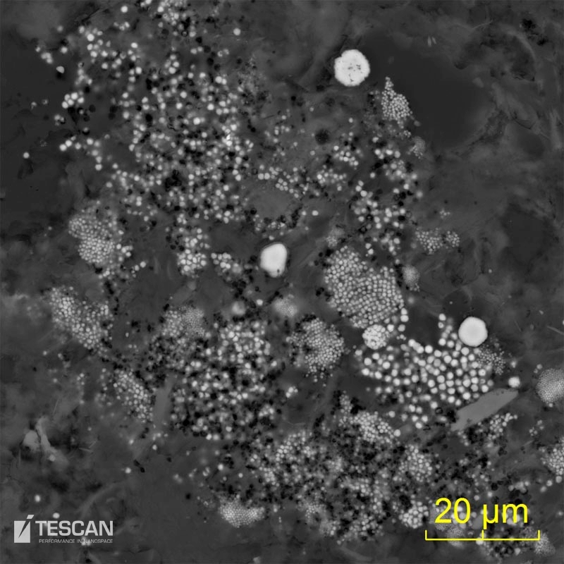
- Framboidal pyrite in a shale rock
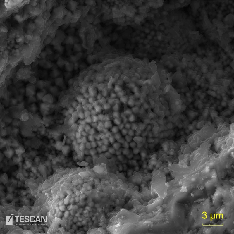
- Pyrite framboid on the crack of coal seam
- Cathodoluminiscence and BSE image of a quartz sandstone with a goethite matrix
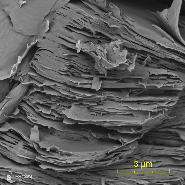
- Clay particles aglomerate in coal


