Quantitative phase imaging (QPI) provided by Q-PHASE, a TESCAN multimodal holographic microscope, allows precise detection and quantification of changes in cell dry mass and related morphological parameters specific to cell death. Thus, individual phases of cell death can be easily distinguished based on the monitoring of a single cell behavior over time. Using the Q-PHASE, significant changes of cellular parameters have been detected in human prostate adenocarcinoma LNCaP cells treated with a chemotherapy drug doxorubicin (DOX).
Do you want to know more information? Please download our Application Example.
If you want to see Q-Phase in practice, you can register here for workshop.
-
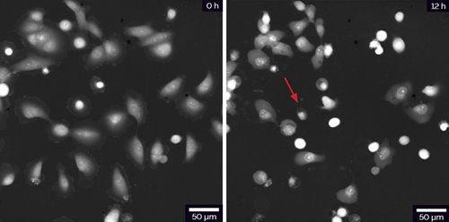
- Changes in morphology of LNCaP cells treated with DOX. Red arrow indicates an example of dead cell in QPI image.
-
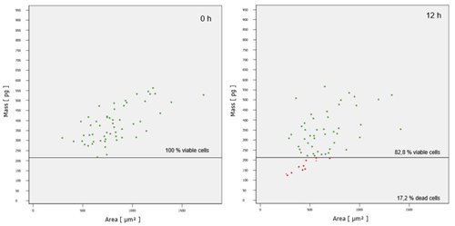
- Cells with small area and dry mass below the determined mass limit (220 pg) are considered dead (red dots).
-
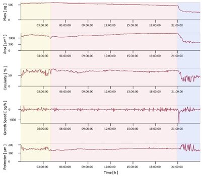
- Changes in morphological and dynamic parameters during necrosis of LNCaP cell. Yellow area denotes an interval of metabolic activity prior to cell swelling shown in red area. Blue area denotes a loss of cell membrane integrity with sheer drop of mass