The structural subcellular analysis provides information hidden under the surface of cells and tissue. Typically this area of research has been a domain of transmission electron microscopy. However, SEM technology is becoming more popular in this field, due to emerging techniques available with scanning electron microscopy solutions. TESCAN offers several solutions for scientists interested in the subcellular investigations of biological samples.
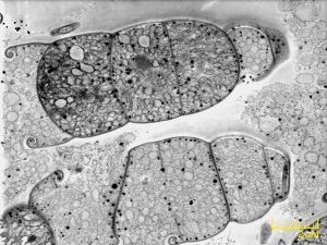
STEM Image of cyanobacteria
- The first approach involves incorporation of a removable STEM detector inside the SEM chamber, thus transforming any TESCAN SEM into a low-kV TEM instrument.
- STEM detector is fully compatible with standard grids routinely used in TEM microscopes.
- Another approach uses serial sectioning inside the SEM instrument followed by imaging. The sectioning can be done either by implementing an ultramicrotome inside the chamber or directly by focused ion beam technology.
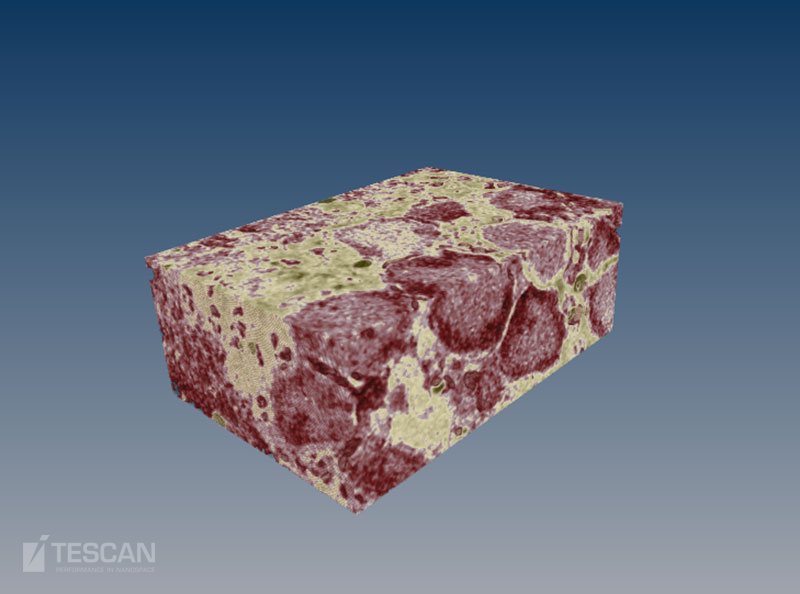
- By serial sectioning of the samples followed by subsequent segmentation and rendering, researchers can easily visualize the relevant data in 3D.
- 3D reconstruction of retina tissue
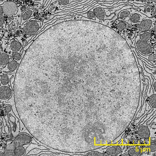
- Serial block phase image of a cellnucleus
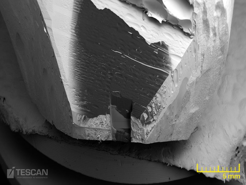
- Large area FIB-SEM cross-section in a resin-embedded tooth
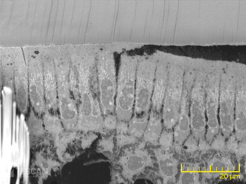
- Osteoblast layer in a mouse tooth


