Non-coated and highly charging samples can be observed either by using low accelerating voltages or under cryogenic conditions. With our dedicated FIB-SEM systems, researchers can cut directly into the materials and investigate material-tissue interfaces or reconstruct them in 3D.
The surface properties and interactions between the material and tissue are keys for predicting the biocompatibility of the material with the host body. This is particularly important in designing reliable implants, tissue mimics and bone replacements, among others. TESCAN SEM systems are ideal instruments for the characterization of biomaterials with high resolution. Wide Field Optics™ allows easy navigation on the sample at very low magnification in real-time.
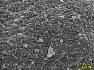
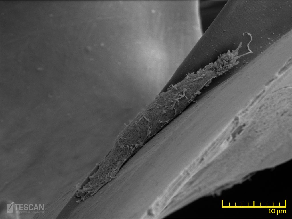
Polymer microfibers with silver nanoparticles
- Once the region of interest is found, the SEM column can be easily switched to a high depth of field or high-resolution modes for detail inspection of the features at high magnification.
- All instruments can be easily equipped with a wide variety of detectors and analytical methods such as EDX and EBSD analysers, Raman spectrometers for detailed compositional and structural analyses of biomaterials.
- Non-coated and highly charging samples can be observed either by using low accelerating voltages or under variable pressure conditions in the UniVac mode. With our dedicated FIB-SEM systems, researchers can cut directly into the materials and investigate material-tissue interfaces or reconstruct them in 3D.
-
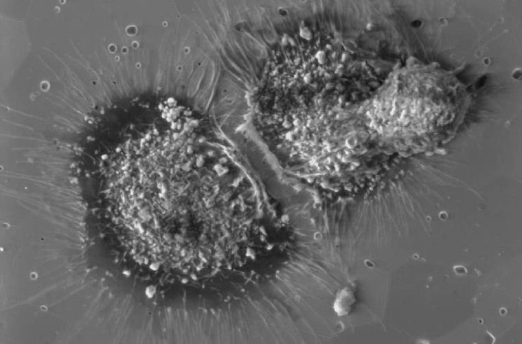
- Spreading of the osteoblasts on the zirconia ceramics
-
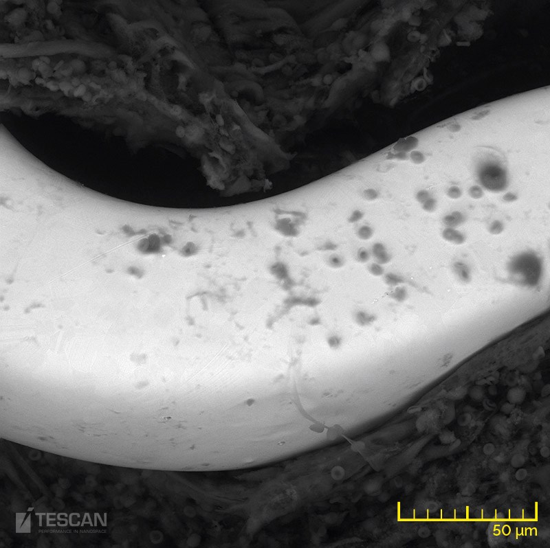
- A piece of cardiac tissue with an implanted cardiovascular stent – BSE detector
-
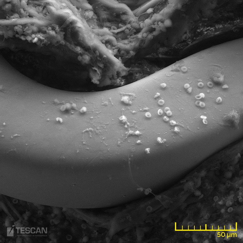
- A piece of cardiac tissue with an implanted cardiovascular stent – LVSTD detector
-
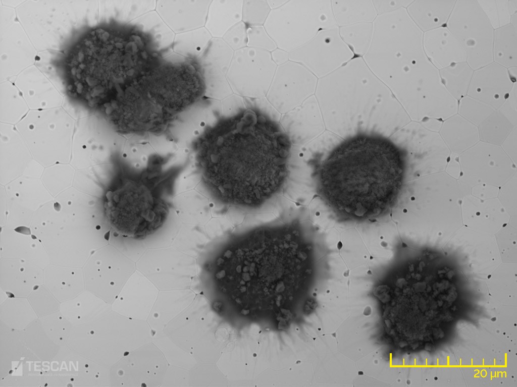
- Growth of osteoblasts on a zirconia ceramics
-
- A fibroblast growing on a collagen scaffold