.png?width=620&height=620&name=GS(1).png)
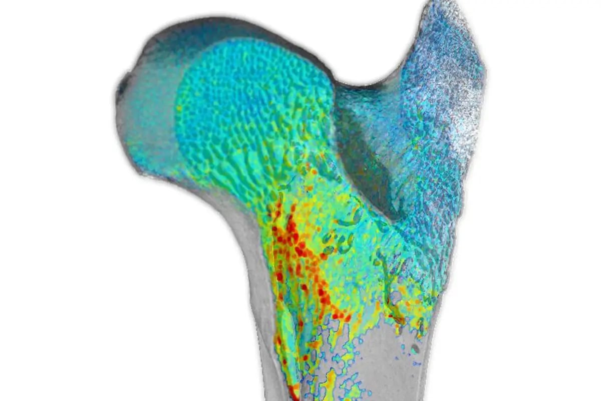
Characterize reservoir rocks and porous media with precision and reliability. Tescan micro-CT workflows visualize connected pore networks and quantify capillary trapping, wettability, and fluid distribution under realistic reservoir conditions.
High-resolution 3D imaging and dynamic pore-scale analysis reveal how CO₂ and H₂ interact with brine, minerals, and rock microstructures. This delivers quantitative data that supports safe, long-term storage validation and reservoir performance modeling.
Gain reproducible pore-scale insights that advance research in geological carbon sequestration, hydrogen storage, and renewable subsurface energy systems.
-2.png?width=433&height=278&name=1_submicron%20scan%20of%20a%20carbonate%20sample%20(Savonni%C3%A8res%20Limestone)-2.png)