TESCAN
menu
en

NEW FRONTIERS OF MICROROBOTICS  
HOW SMALL CAN WE GO?

NEW FRONTIERS OF MICROROBOTICS  
HOW SMALL CAN WE GO?

Did you know that robots are now being developed that are so small you need a microscope to see them? Known as “microrobots,” these micro-scale robots have the potential to revolutionize health care and more. 

When we think of robotics, we tend to think of the massive machines that make cars, or the arms and legs of amputees, maybe even the humanoid creations of science fiction. But robots are now being developed that are so small you need a microscope to see them. 

Figure 1: The microgripper is printed above the 5 μm diameter core of the 125 μm diameter single-mode optical fiber. Light from a laser is shone through the fiber, and the reflected spectrum can be read back. Thin layers of polymer (which are approximately 1 μm thick) are suspended by 3 springs and these give rise to an interference pattern that is reflected back. As the gripper closes, the air gaps between the polymer layers decrease and the interference pattern also changes as a result. This fact can be used to train a model to learn how to estimate forces on the gripper using reflected spectra.

These microrobots have the potential to revolutionize health care, for example, by replacing invasive, often complicated surgery with less invasive techniques. To date, most of the research has focused on autonomous, swimming-type microrobots, propelled and controlled by magnetic fields, Sometimes by ultrasound or light. However, microrobots can be tethered with the advantage that the connection can deliver a power source as well as control the microrobot’s global position. 

Recently, a research team at Imperial College London reported the development of a tethered microrobot, a microgripper that is no bigger than 100 µm on each axis. Remember that a µm, or micrometer, is one-millionth of a meter! It is so small that you need a scanning electron microscope to see what the gripper (Figure 1)

looks like and to monitor its construction and operation, in this case TESCAN microscope. The research team used a relatively new 3D printing process, 2-photon polymerization, to fabricate the microgripper. Ultra-short laser pulses trigger a highly localized chemical reaction in a volume of soluble photosensitive material similar to that used in  semiconductor manufacturing. Once exposed, the material becomes insoluble and, after washing in a developer bath, a self-supporting 3D structure is left behind. This process allows fabrication of microscale structures as small as 100 nanometres – that is one-tenth of a micrometer! 

Using this technique the team fabricated the microgripper – in a single process, with all its parts assembled. They also printed it directly onto the tip of a 125 µm diameter optical fiber that carries only a single ray of light. Tethering the gripper in this way might sound simple, but this is the first time that such a small fiber-based micro-robotic system has been developed.  

HOW DOES THE MICROGRIPPER WORK?

A robotic gripper has three main features. Firstly, the device needs a control system to enable it to navigate and position itself around its environment (global actuation).

This was straightforward and achieved by a commercially available micromanipulator that translates large-scale hand movements into micro-movements, in this case using a joystick or autonomously by a computer. Secondly, gripping was achieved by relative motion along the tool’s central axis which resulted in the gripper’s fingers closing (local actuation). Finally, the gripper needs to sense the force (force-sensor) and provide feedback to determine subsequent actions. In a rare achievement, the research team combined the gripping and feedback capabilities (local actuation and sensing) into a single microrobot. The novel design of the microgripper is shown in Figure 2.

It has three equidistant fingers joined together at the base by a central ‘palm’. Attached to the palm and to the base of the fingers are three springs. These hold four parallel thin plates in place. When the gripper touches the object to be grasped a force is applied to the palm, the hinges move, the fingers close inwards, the springs compress, and the distance between the parallel plates reduces. This movement of the parallel plates is the key to the microgripper’s force-sensing mechanism. 

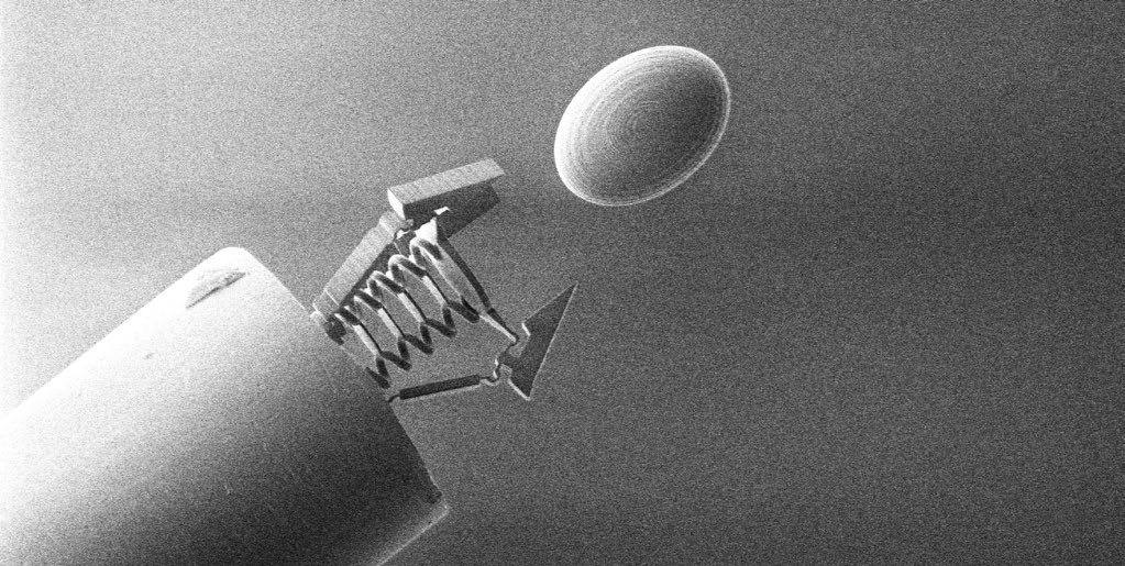
Figure 2: A microgripper, printed using 2-photon polymerization (a microscale 3D printing technique) onto an optical fiber substrate, approaches an ellipsoidal object. This rapid-prototyping fabrication method makes it easy to tailor the geometry and size of the micro tool to suit the target object. The gripper is a passive, compliant design with three fingers arranged 120° from each other.

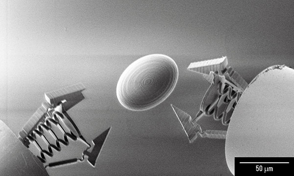
Figure 3: Two similar microgrippers approach a target object. Using two such grippers, the target object
can be manipulated or transported in a cooperative manner with real-time force readings.

Supercontinuum light, best described as ‘broad as a lamp but bright as a laser,’ is directed through the tethered optical fiber and is partially transmitted and partially reflected by each of the parallel plates. Changes in the relative distances between layers of plates result in different reflected light spectra. This is collected by the optical fiber and delivered to a spectrometer. Analysis of the spectral light readings provides a measurement of the compression state of the gripper. The principle of the optical force-sensing is shown in Figure 3. The research team calibrated the microgripper using the spectral light readings to measure the applied force and then proceeded to demonstrate that the microgripper could be used to hold and move objects in a reliable and repeatable manner.

WHERE TO FROM HERE?

Over the last few decades a variety of general purpose, electromechanical, microscopic grippers have been produced using different fabrication methods, different methods of actuation and different sensory mechanisms. However, this is the smallest tethered force-sensing microgripper ever manufactured and world’s first ever force-sensing micromanipulation tool fabricated by the 2-photon polymerization method. The device holds significant potential for use in cellular manipulation 

and microscale surgery as it is capable of manipulating soft and fragile objects at the cellular level. It is also easy to manufacture. Furthermore, although it is a passive system in that the fingers close only 

when the ‘palm’ is touched and the force applied, the optical fiber-based design has the potential to deliver energy to activate the gripper (full local actuation). The research team at Imperial College will explore this in future designs to bring the dream of cellular-level microsurgery that little bit closer. TESCAN’s scanning electron microscopes will continue to play an essential role in enabling these developments. 

NEW POSSIBILITIES WITH BOTTOM-UP MANUFACTURING

The advent of bottom-up manufacturing including 3D printing processes has provided scientists with a new level of freedom to be able to tailor make microscopic robots from the ground up. This has allowed them to fabricate smaller robots than conventional top-down methodologies. These robots have the potential to treat diseases at the cellular level and to perform surgeries in even less invasive ways than is currently possible. 

MAURA POWER

Paul Wood and Maura Power from Imperial College London


recently completed her Ph.D. at Imperial College London Developing novel micro robotic grippers 

with sensing and actuation capabilities, and is currently a post-doctoral researcher working towards the clinical translation of these microscale tethered tools. This work is part of the larger “Fibrebot” project for endoluminal surgery. Her interests are in the design, fabrication and characterization of devices that could, in the future, provide surgeons with new tools and capabilities. She is also interested in the automation of experimental data acquisition.


13.04.2021