Tescan SEM (Scanning Electron Microscope) systems deliver high-resolution surface imaging and analytical versatility for research, industrial quality control, and failure analysis across a broad range of applications.
Combining advanced electron optics with intuitive workflows and flexible analytical integration, Tescan SEM platforms enable users to investigate materials and structures from the microscale down to the nanoscale with speed, precision, and confidence.
Built on decades of expertise in electron microscopy and charged particle optics, the Tescan SEM portfolio supports applications spanning materials science, semiconductors, battery research, metallurgy, geology, life sciences, and additive manufacturing. From routine imaging and particle analysis to demanding low-voltage characterization and in situ experimentation, Tescan SEM solutions are designed to adapt to evolving scientific and industrial challenges.
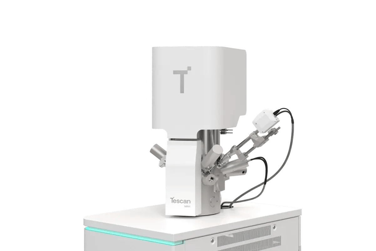
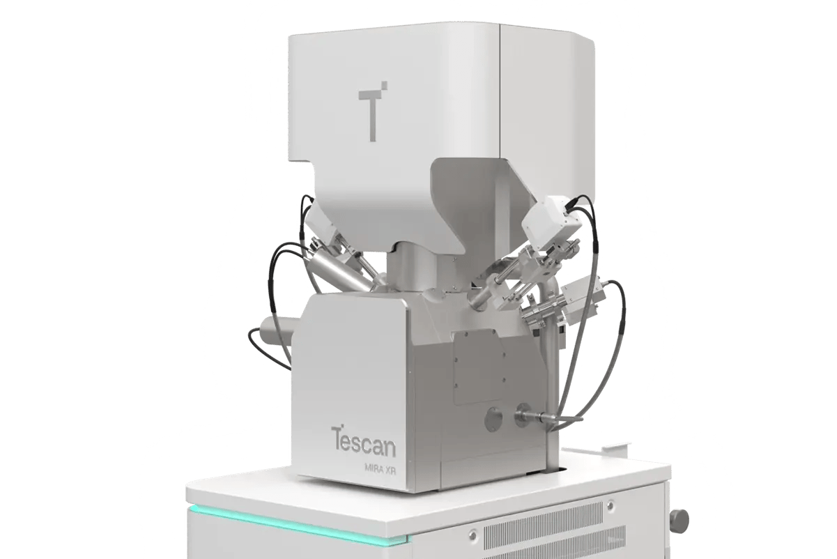
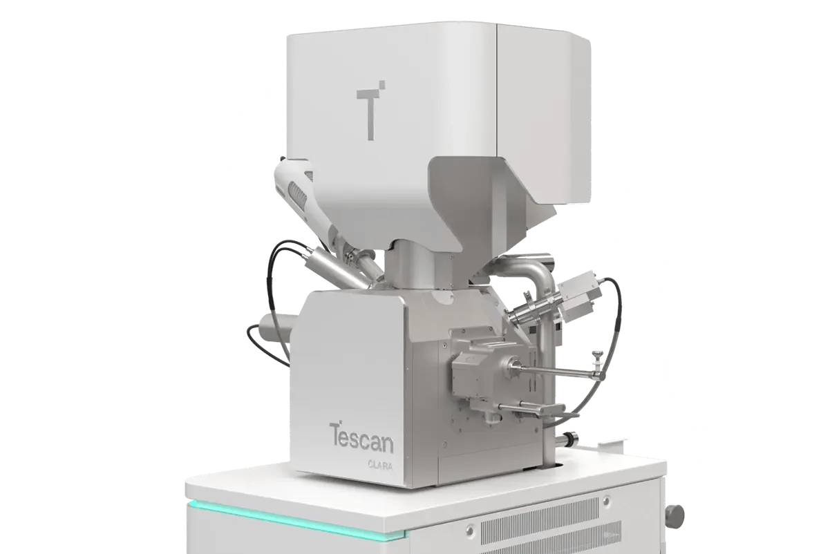
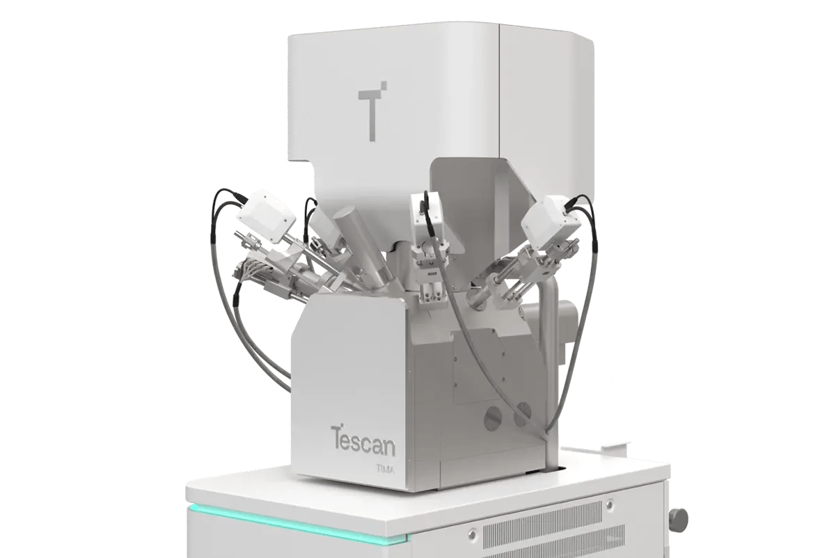
The portfolio includes thermionic SEM systems for accessible and reliable routine analysis, as well as advanced field emission SEM (FE-SEM) platforms optimized for ultra-high-resolution imaging, surface sensitivity, and analytical performance. Field-free imaging technologies, advanced detection systems, and low-energy beam capabilities allow users to examine beam-sensitive, non-conductive, and complex materials while preserving sample integrity and maximizing image quality.
Tescan SEM systems support a wide ecosystem of analytical and correlative workflows, including EDS, EBSD, WDS, Raman, cathodoluminescence, tomography, and automated image analysis. Modular architectures and open platform design enable laboratories to configure systems around their specific applications while maintaining flexibility for future expansion.
Not sure which SEM configuration best fits your application, workflow, or throughput requirements? Request a personalized consultation with a Tescan specialist to discuss your samples, analytical goals, and laboratory needs, and identify the solution best suited to your research or production environment.


.webp?width=1072&height=741&name=VEGA%20Compact%20(3).webp)