Join our "Opening Seminar" and extend your knowledge of the most innovative technologies in cryo-electron microscopy
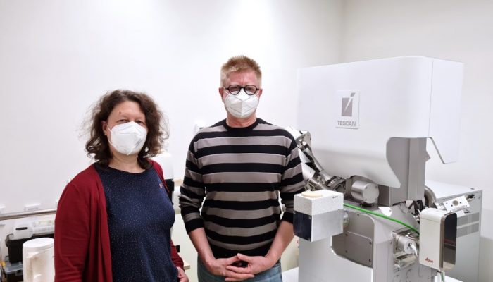
Institute of Molecular Genetics of the Czech Academy of Sciences (IMG) together with industrial partners TESCAN and Leica Microsystems is organizing an Opening Seminar of their collaborative project.
This free on-line seminar will take place on 26 May 2021 from 10:00 to 12:00 AM CEST.
PROGRAM
May 26, 2021
10:00 – 10:10
Prof. Pavel Hozák (Institute of Molecular Genetics, Prague)
- Imaging possibilities under Czech BioImaging umbrella
- Introduction of the collaborative project between IMG – TESCAN – Leica
10:10 – 10:45
Dr. Vlada Filimonenko (Institute of Molecular Genetics, Prague)
- Study of lipidic structures in the cell nucleus by means of electron microscopy
- Opportunities for your research at the Electron Microscopy Core Facility IMG
10:45 – 11:25
Dr. Ondřej Šulák (TESCAN Orsay Holding a.s.)
- TESCAN AMBER cryo – Versatile FIB-SEM solution for cryo and room temperature
structural analysis
11:25 – 12:00
Dr. Frédéric Leroux (Leica Mikrosysteme GmbH)
- Workflows and instrumentation for cryo-electron microscopy
12:00 – 12:05
Prof. Pavel Hozák (Institute of Molecular Genetics, Prague)
- conclusion, end of the seminar
The IMG EM CF is supported by MEYS CR (LM2018129, CZ.02.1.01/0.0/0.0/18_046/0016045, CZ.02.1.01/0.0/0.0/16_013/0001775).
Study of lipidic structures in the cell nucleus by means of electron microscopy
Opportunities for your research at the Electron Microscopy Core Facility IMG
Presented by Vlada Filimonenko, Head of the Electron Microscopy Core Facility at IMG
The interior of the nucleus of eukaryotic cell has highly organized architecture that allows to complete all specific functions, such as gene expression, DNA repair or RNA processing. Our laboratory has recently identified the novel membrane free nuclear Phosphatidylinositol 4,5 bisphosphate (PIP2) containing structures – Nuclear Lipid Islets (NLIs) that play important role in modulation of transcription by RNA Polymerase II. High resolution spatial characterization of the NLI and other lipidic structures in the cell nucleus upon various experimental conditions is crucial for understanding their important roles. To perform observations on a sample as closely as possible to the living state, the best way is cryo-immobilization and imaging under cryo-conditions. Fabrication of a thin FIB-milled lamellae from a frozen hydrated sample with subsequent cryo-transfer and tilt series acquisition in cryoTEM is currently the best workflow introducing minimal artefacts into the sample compared to other available techniques. The workflow is technically challenging and needs significant skills and optimization of all steps to produce homogeneously thin lamella and to avoid heat damage, mechanical damage, and surface contamination of the lamella. The whole workflow is being implemented on the basis of the Core Facility for Electron Microscopy at IMG. Being a part of the Prague Node of the Czech-BioImagning and EuroBioImaging infrastructures, the core facility provides open access to a broad range of technologies in the field of transmission electron microscopy and biological sample preparation.
About Vlada Filimonenko
Dr. Vlada Filimonenko is the Head of the Electron Microscopy Core Facility at IMG and a scientist in the Department of Biology of the Cell Nucleus IMG CAS headed by Prof. Hozák. Her main research interests lie in the field of relationship between structure and function of the cell nucleus and the principles of formation and maintenance of nuclear functional compartments.
TESCAN AMBER cryo - Versatile FIB-SEM solution for cryo and room temperature structural analysis
Presented by Ondřej Šulák, Product Marketing Director – Life Sciences
Low-temperature electron microscopy (cryo-EM) has become an established technique for capturing and observing beam-sensitive samples in their close-to-natural state.
Cryo-sectioning is a standard method used for thinning and slicing such samples. However, the integrity of the specimens can be degraded easily by common artifacts such as knife marks, compression, or crevasses. Focused ion beam (FIB), on the other hand, is used extensively to reveal sub-surface information from bulk material, create samples for other analytical modalities, or prepare ultra-thin specimens for analysis in transmission electron microscopy (TEM), without knife-sectioning artifacts.
Moreover, combining the site-specific milling capabilities of FIB with scanning electron microscopy (SEM) opens a broad range of possibilities for preparing biological samples. FIB-SEM systems are widely used not only for routine preparation of ultra-thin TEM specimens but also for precision cross-sectioning and 3D volume imaging, which are possible in ambient temperatures as well as in cryogenic conditions.
The seminar aims to demonstrate the feasibility and discuss the potential of TESCAN cryo-FIB-SEM as a reliable tool for cryo-TEM sample preparation, with its exceptional range of additional capabilities—all within a single versatile workstation.
About Ondrej Sulak
Ondřej Šulák received his Master’s degree in Analytical chemistry at Masaryk University in the Czech Republic. Then, he moved to France where he continued his Ph.D. studies and Postdoctoral research at Joseph Fourier University in Grenoble where he was focused on the structure-functional characterization of carbohydrate-binding proteins, mainly by X-ray crystallography, Electron Microscopy, SAXS, and other techniques. His industrial career started in the biotechnological company where he was responsible for the biochemistry department. In 2016, Ondrej joined TESCAN as Global Applications Director. Since January 2019 he works as Product Marketing Director for the Life Sciences segment.
Workflows and instrumentation for cryo-electron microscopy
Presented by Frédéric Leroux, Advanced workflow specialist, Leica Microsystems
Cryo-electron microscopy is an increasingly popular modality to study the structures of macromolecular complexes and has enabled numerous new insights in cell biology. In recent years, cryo-electron microscopy has expanded further towards in situ structural biology and has become the go-to technique for resolving structures in their native context.
Similarly, freeze-fracturing, cryo-scanning electron microscopy (SEM) and cryo-focus ion beam (FIB) have received increased interest. Cryo workflows typically start with vitrification, and all subsequent steps—including screening, processing, transfer, and imaging—are performed under cryogenic conditions.
During this presentation, you will receive a full overview of cryo-preparation instruments, including vitrification, light microscopy screening, sectioning, planning, fracturing, milling, coating, and transfer. All of these steps happen under cryogenic conditions while minimizing contamination and without risk of devitrification.
About Frederic Leroux
Frédéric Leroux completed his Master degree in Biology in 2007 at the University of Ghent where he gained experience in biological EM sample preparation. In 2008, he moved to the physics department at the University of Antwerp where he started his PhD. At the EMAT research group he specialized in advanced electron microscopy of composite materials. 2016, he joined Leica Microsystems as Application Specialist Nanotechnology EMEA.
He received his PhD in 2012. After 2 years as a postdoctoral researcher he became EM sample preparation specialist at EMAT. He thereby uses his multidisciplinary background and broad microscopy experience to improve EM sample preparation of a variety of materials (polymers, composites, biological and industrial materials).
