In addition to its structural role, a membrane is also a place, where many important events occur, such as cell differentiation, signalling and interactions with extracellular space. Changes in the morphology of the outer membrane can be evidence of altered cellular functions, such as tumour formation, cell-pathogen interactions and stem cell differentiation. Typical applications involve observing shape changes of grooves, pores, blebs or microvilli on the cellular in response to the changes in the extracellular environment. Delicate samples can be observed in non-dehydrating conditions in under elevated pressure and increased humidity using the UniVac mode and the Water Vapor Inlet.
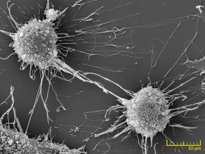
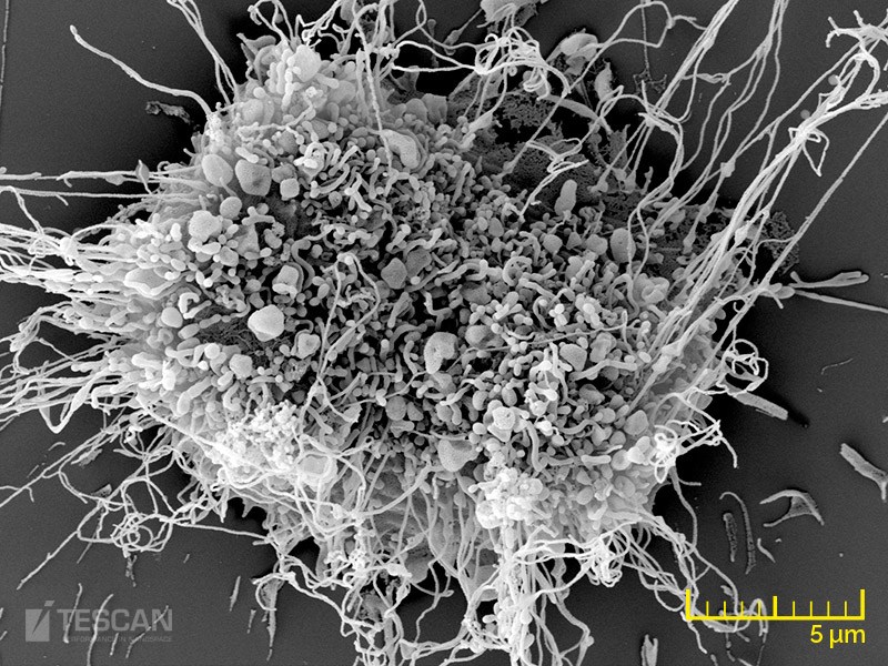
Fibroblast culture
- TESCAN MIRA3 and MAIA3 FEG-SEMs are ideal instruments for investigating cellular and tissue structure with high resolution.
- The MAIA3 is specifically designed for ultra-high resolution imaging at low accelerating voltages, thus providing the best topographic contrast and detail.
- Detail of a fibroblast cell
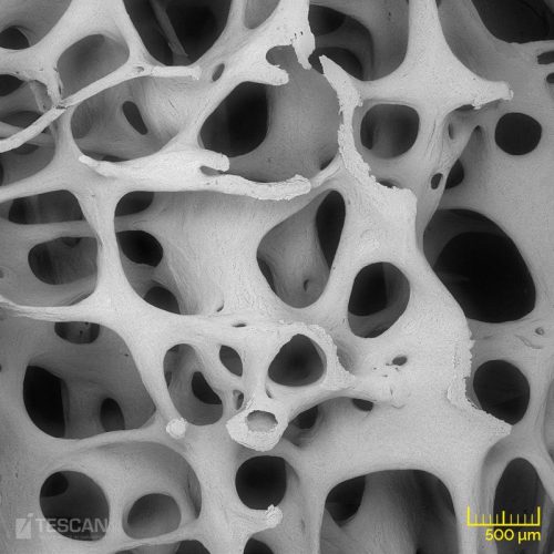
- Rat bone structure
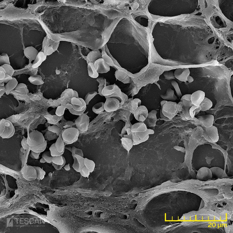
- Adipose tissue
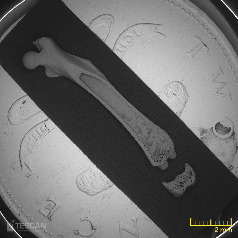
- Whole rat femur


