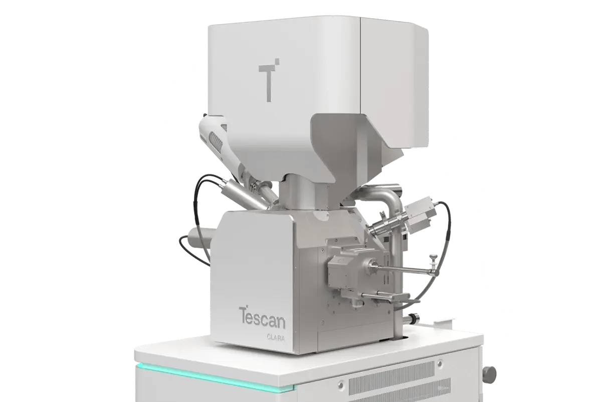
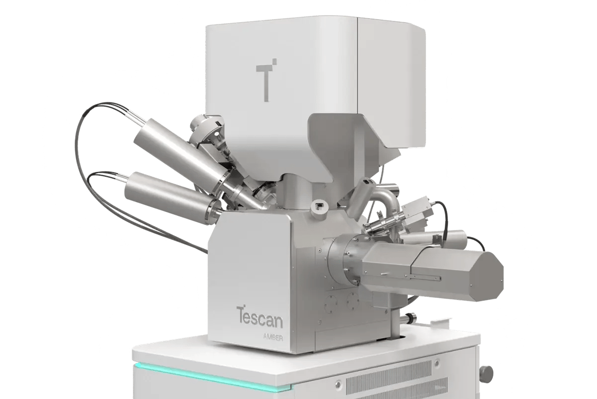
TESCAN
menu
en

Automated time-lapse segmentation by TESCAN Q-PHASE

Read the difference among methods for label-free light microscopy imaging of cells

Automated time-lapse segmentation by TESCAN Q-PHASE

Phase Contrast (PhC) and Differential Interference Contrast (DIC) are the most traditional and commonly used methods for label-free light microscopy imaging of cells. Both methods are based on the fact, that light passing through the cell increases phase when compared to the background. But neither PhC nor DIC provides the quantitative information about this phase increase.

Although from the DIC and PhC images it is easy to recognize the cell details by naked eye, the process of automatic segmentation made on such images is much harder. Some of the modern segmentation methods which work with DIC and PhC images try to first recalculate captured data into Quantitative Phase Image (QPI) data, as QPI data are extremely suitable for segmentation due to the good separation between background (zero values) and cells (non-zero values). Anyway, such recalculation of QPI is not precise. Obtaining the QPI information directly would make the process of computer segmentation much easier and reliable.

TESCAN Q-PHASE microscope provides QPI information directly. Moreover, thanks to the unique patented setup it also enables to use incoherent light, thus creating high quality images without any coherence artefacts. Therefore Q-PHASE is extremely well suited for precise time-lapse segmentation which is made automatically using specially designed Q-PHASE software.


20.02.2017