In this application example, we demonstrate by using the new TESCAN S9000G microscope, that FIB-SEM technology is an innovative approach to study and resolve the complexity of subcellular environment. The advancement in 3D imaging allows the precise morphological description of individual organelles and identification of their mutual connections. Given the reliability, efficiency, and high resolution of FIB-SEM technology, this analysis represents an important foundation to better understand organelle cross-talk in mammalian cells.
Would you like to know more information? Download our Application Example.
-
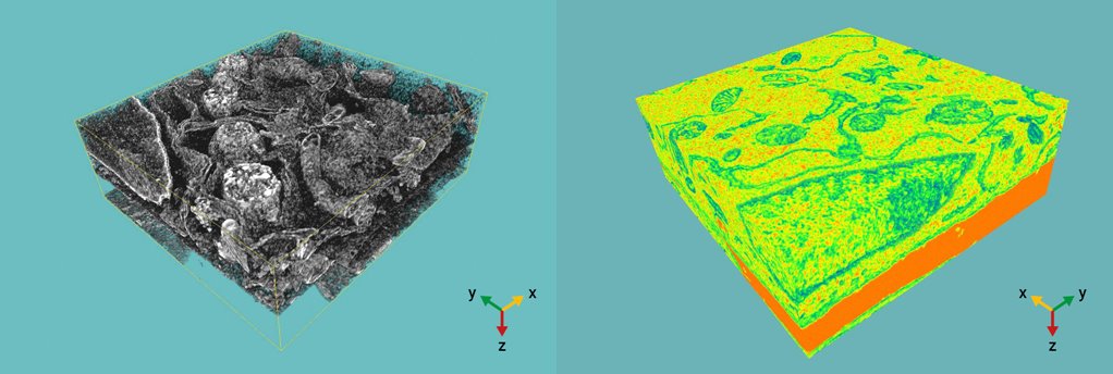
- 3D ultrastructural reconstruction of studied mammalian cell. Selected FIB-SEM stacks show intracellular organization with clear contacts between organelles.