Somatostatinoma is a type of a neuroendocrine tumor that typically develops in the region of the duodenum or pancreas. Elevated levels of somatostatin secreted can be used to investigate somatostatin tumor cells, typically by the positive immunohistochemical staining. Correlative light-electron microscopy imaging has become a very popular approach due to the usage of quantum-dot (QD) based immunoprobes, which can be inherently detected by both techniques.
The TESCAN MAIA3, an ultra-high resolution SEM is capable of detecting QD-immunolabeled sections prepared on glass slides (typical size of QDs is below 10 nm). The TESCAN MAIA3 offers excellent performance at low beam energies thanks to its Triglav™ SEM column technology that is equipped with an advanced detection system which provides with compositional contrast and allows low-noise volume compositional mapping.
Download the new TESCAN Application Example and find out more on this.
-
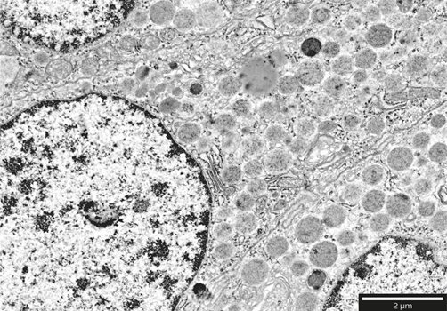
- Localization of secretory granules of somatostatinoma tumor cells in pancreatic tissues. Images acquired at 2 keV.
-
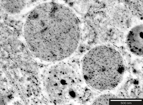
- Localization of secretory granules of somatostatinoma tumor cells in pancreatic tissues. Images acquired at 2 keV.