Palaeontologists typically use secondary electron (SE) observation to reveal the morphology of fossils. Secondary electrons are formed very close to the sample surface, have a high spatial resolution and reveal very fine details of the sample surface. They also have a larger depth of focus than light microscopy. Electron microscopes are capable of resolving details at the sub-nanometre range compared to hundreds of nanometres in optical systems. Higher depth of focus is needed for imaging large three-dimensional complex-shaped fossils and fossil remnants.
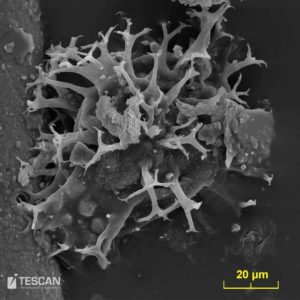
Dynoflagelata
- Electron microscopy is typically used in palaeontology for the identification and taxonomy on microfossils. A conductive coating is usually applied to discharge the electron beam current.
- Microfossils are generally smaller than a few millimetres. The most common organisms are algae, protozoa, and crustaceans. Large populations of microorganisms occurred over a very wide geographical area. Their wide distribution and relative sensitivity to environmental changes have made microfossils into so-called index fossils that are used in the dating of sedimentary sequences.
- Macrofossils can also be studied because of the high depth of focus. Similarly, the bones of small vertebrates can also be studied to determine the causes of different surface marks.
- Specimens which cannot be coated by a conductive layer can be observed under the low vacuum conditions preventing the accumulation of charge.
- A special SE detector was developed for this purpose – LVSTD (Low Vacuum Secondary Electron TESCAN Detector).
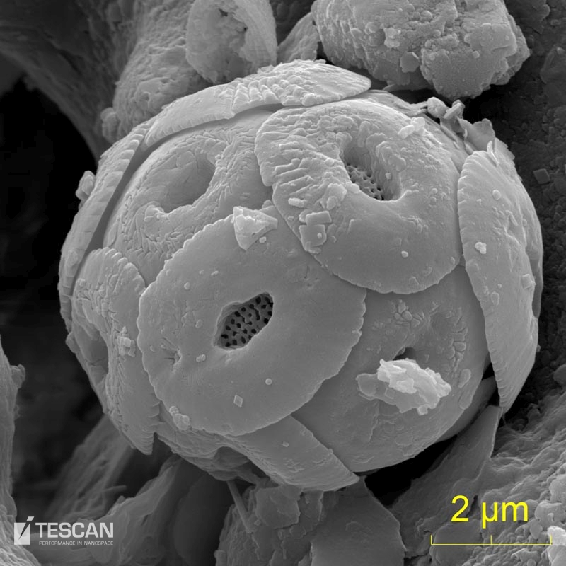
- Coccolithophore
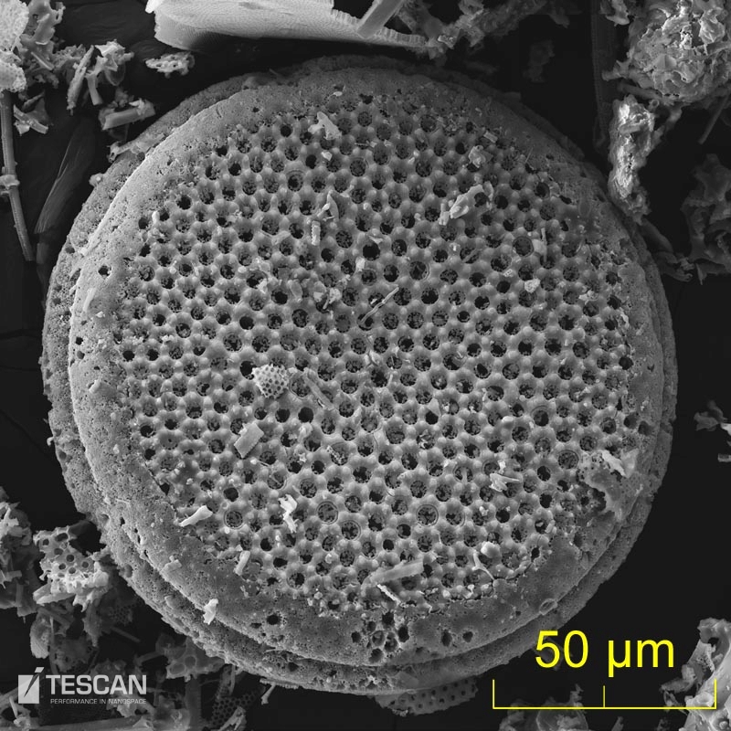
- Diatoms
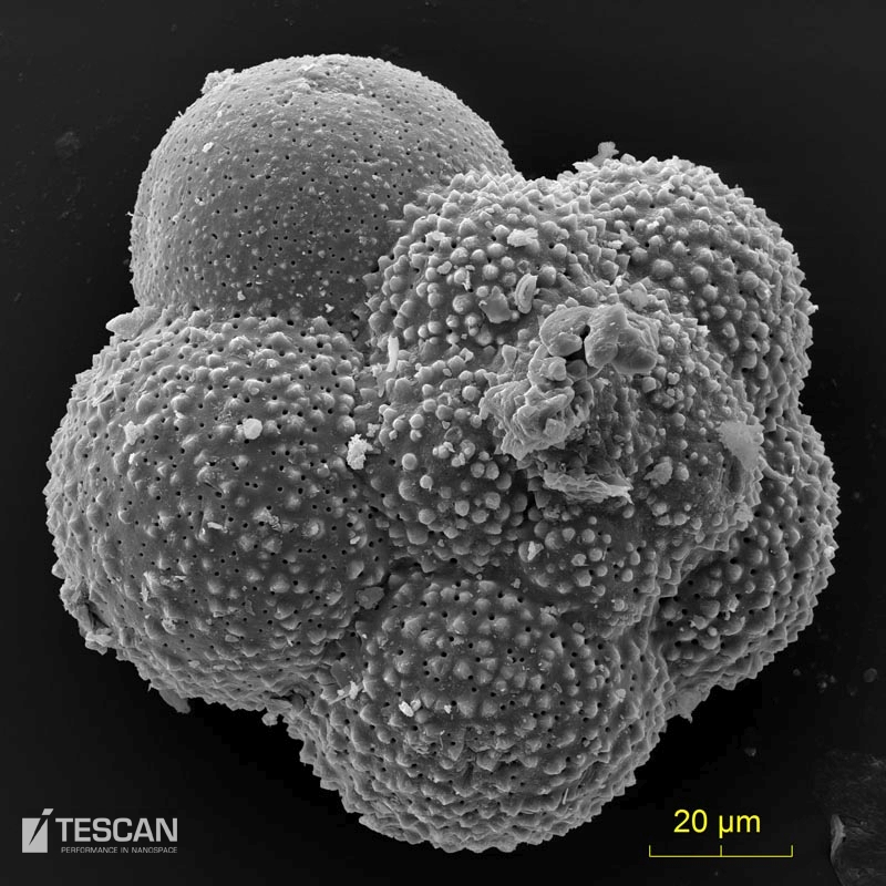
- Foraminifera
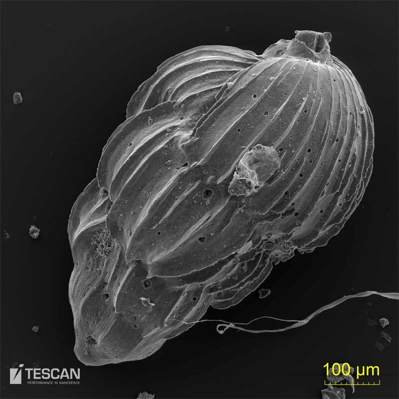
- Foraminifera – uvigerina


